As energy geoscientists we continually strive to understand the rocks that are the building blocks of energy systems. Whether that be in the hydrocarbon systems that have underpinned industrial development over the last two hundred years, or in those systems that are key to the energy transition as society shifts away from fossil fuels, such as geothermal, heat and energy generation and storage (e.g., hydrogen, compressed air) and also long-term storage and disposal of CO₂ and radioactive waste.
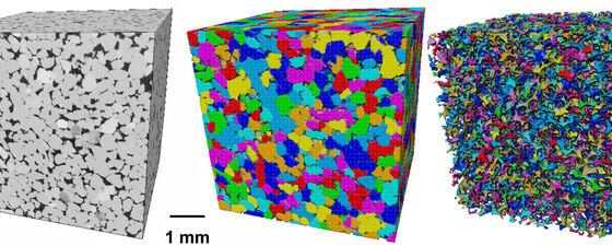
3D image of sandstone, resolving grains and pore-networks (part of an ongoing CCS-reservoir potential study). © Taylor and Ma.
Modeling is Key
Whilst the large-scale (e.g. geophysical) and traditional petrographic techniques have continued to evolve over the last three decades or so, the last ten years has also seen enormous growth and development in the application of new X-ray and electron microscopy techniques to image, in three and four dimensions, the microstructure and nanostructure of rocks (both porous reservoirs and relatively impermeable seals) in order to better understand sealing properties, fluid/gas storage and flow in these rocks, as well as to build physical and chemical models to effectively simulate and predict subsurface behaviour. All subsurface energy technologies depend upon fluid movement and storage as well as impermeable cap rocks to retain the fluids in the subsurface. In order to do this effectively, and understand key uncertainties and manage the resultant risks, there is a critical need to understand in both 3D and 4D wherever feasible, porosity (fluid storage), porosity connectivity (permeability) together with microfracture characteristics (transmissibility and seal integrity). Whilst offering significant opportunities to understand these systems with new techniques and technologies being developed and improved all the time, there are still challenges and workflows that need to be understood in applying these techniques.
It’s a Matter of Scale
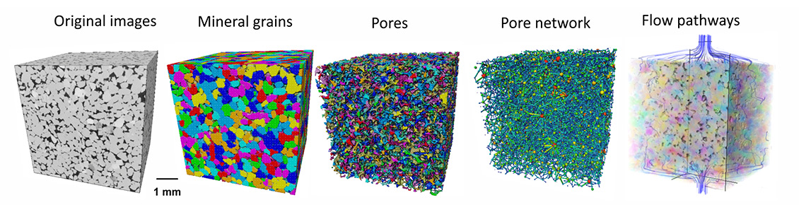
There are many techniques that can be applied; these are selected based on the resolution required, and what can be achieved by each and on the features that we wish to image and quantify. As can be seen from Figure 1, the scale and resolution of interest dictates the method selected. Macro-X-ray Computed Tomography (CT), micro-Xray CT, nano-X-ray CT, Focussed Ion Beam Scanning Electron Microscopy (FIB-SEM) and Transmission Electron Microscopy (TEM) tomography, are techniques that, respectively, offer a sequence of increasingly higher spatial resolution. In the workflows we apply, it is important to be aware that as imaging resolution improves, and smaller features can therefore be resolved (in effect the voxel size – a 3D pixel – decreases), the volume size of rock that can be imaged also gets proportionately smaller. At the extreme end, the volume of sample that can be imaged by TEM tomography is as small as tens of nanometres (nm); this offers a resolution as small as 0.1 nm. At the other end, microfocus, X-ray tomography (macro-XCT) can image very large samples such as core material, but image resolution is rarely better than a few millimetres. It follows that we must select carefully the imaging method based upon the features we are interested in studying. For sandstones and other coarser granular materials, we would primarily be looking to use microfocus X-ray tomography (micro-XCT) for pore imaging, whereas for microfractures in cores/mudstones/carbonates, macro-XCT is commonly good enough. For pores in shales, where high resolution is demanded, then the techniques of FIB-SEM and TEM tomography are needed to understand fully the nature of the rocks.

Figure 1: Multiscale methods for 3D imaging, characterisation and modelling. © Taylor and Ma.
Challenges and Opportunities
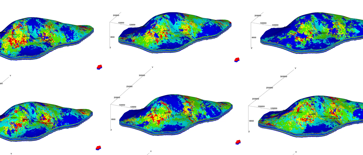
The obvious question arises, whether the images and features that are measured in a sample are representative of the rock as a whole and how can we upscale observations whilst retaining the integrity of the original observation or measurement? This is a difficult challenge that people have addressed in two ways. The first way is to collect multiple images at the same resolution and scale and stitch them together into a larger 3D volume – a 3D version of the widely used gigapan approach in 2D. This results in a larger volume of imaged rock, but at a high spatial resolution. Such an approach is often time-consuming and costly, as well as generating very large datasets; with the advent of machine learning this is more achievable and offers a more viable option than in the past. The other approach is to develop mathematical upscaling models that can be applied to datasets. The approaches for this have been varied and taken the form of several different mathematical solutions; here a number of robust methods have recently been developed and published that allow for increased confidence in the representativeness of the final result.
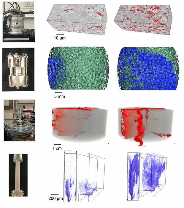
Figure 2: 4D imaging examples of thermal expansion, stimulated fracturing, multi-phase flows and mineral reactions. © Taylor and Ma.
An exciting recent development has been the application of 3D imaging techniques to show the evolution of micro- and nanostructures under subsurface-realistic conditions often with the application of external factors such as heat, pressure and chemical fluids. This has come about with the development of higher flux lab-source X-ray tomography and increased flux and coherent X-rays from new generation synchrotron facilities. These advances in techniques have made taking a series of 3D images over time (i.e., 4D imaging) possible. The time resolution of 3D images depends on the CT scanners and scales. Typically, it takes a few hours to acquire a 3D tomographic image using a laboratory-source X-ray CT scanner and so reactions and changes in rock microstructure and fluids over hours or days can be captured by using this technique. For more rapid changes in samples, such as fracture propagation in response to pressure or heat stress changes, synchrotron-source X-ray CT, that can collect a full 3D image in seconds, is required to capture the evolution and impact of these processes (see Figure 2).
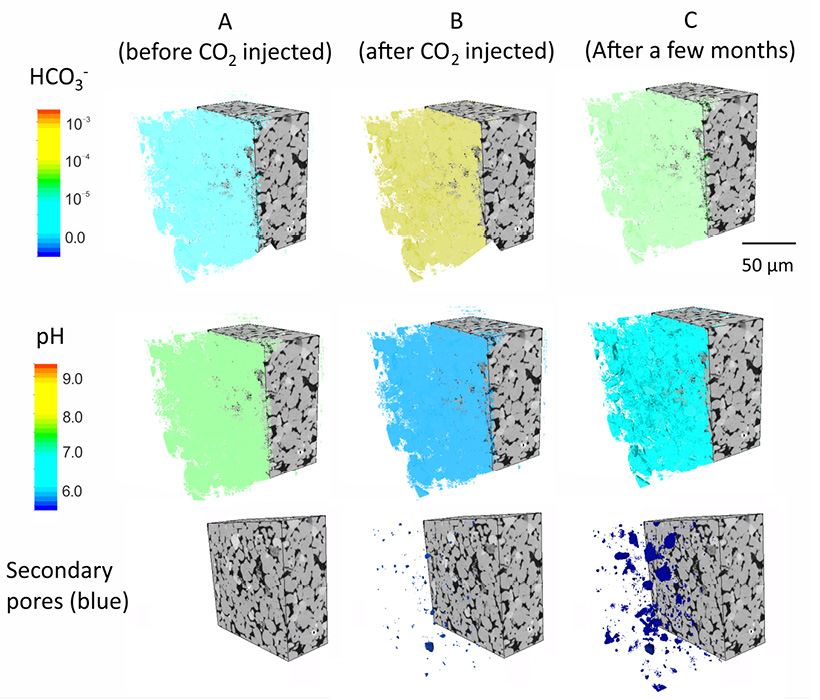
Figure 3: Image-based modelling of carbonate dissolution after CO2 injection in the subsurface. © Taylor and Ma.
Fast reactions, such as multiphase flows, mineral precipitation and dissolution, can also be imaged, quantified and modelled in synchrotron beamlines (see Figure 3). In addition to being able to capture images in 4D, a key challenge is whether we can design rigs to safely confine samples under certain conditions in order to observe the reactions or structural and textural changes that may occur. Researchers have shown that 4D imaging under subsurface conditions, involving temperature (-100 to over 1000°C), pressure (up to 20 MPa), fluids (gas, liquid, supercritical fluids), and chemical conditions (brine, acid, etc) can indeed be achieved using specifically designed rigs (see Figure 2). These rigs allow for samples to be positioned within the X-ray beam and images collected in real time during in-situ experiments. A major limitation of this approach is that in order to
obtain high pressure and/or corrosive fluids, material that would normally be used to contain these conditions such as steel, is not transparent to X-rays, and thus there are limitations to what can be analysed. The development and increasing application of neutron-scattering and neutron-imaging techniques, where steel and other metals are neutron-transparent, will likely lead to further advances in this field.
New Techniques Allow Observation of Fast Reactions
3D and 4D multiscale imaging has proven to have been a very powerful tool in the geo-energy field, both in the academic and industrial sectors. But as discussed above, there are several challenges that we need to address to be able to fully unlock the potential of these 3D imaging techniques, but through careful observational and experimental design, combined with appropriate modelling, these can be solved (see Figure 4).
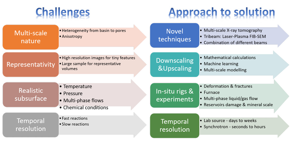
Figure 4: Challenges identified in the 3D and 4D imaging applications and the proposed approach to solutions. © Taylor and Ma.
The application of novel techniques will allow for enhanced observation of heterogeneous features and fast reactions which could not be observed previously. For example, the tri-beam system (laser-plasma-ions FIB) provides both higher resolution and large sample volume (nm-scale static 3D SEM images of mm3-sized sample) to bridge mm- to nm- scales, and the new generation of synchrotron sources, can capture super-fast reactions such as fracturing (down to seconds) through tomography images at high resolution (down to 30 nm). Also, the involvement of machine learning has reduced the experimental and computing time significantly and also provides opportunities to reveal hidden mechanisms behind the images. All of these techniques will further increase the accuracy of modelling (e.g., flows, mechanical, chemical and combination) to improve the theoretical understanding of the rock reactions under complex subsurface conditions and provide optimising suggestions for industrial applications.
In many energy transition applications, the reactions of rock and fluids in the subsurface are poorly constrained. For example, chemical and mechanical reactions during thermal fluids injections for heat storage, hydrogen or compressed air injection for energy storage, and long-term carbon storage and sequestration. Multiscale 3D and 4D image workflows, combined with image-based modelling, will certainly help us open a new chapter for a lower carbon geo-energy future.