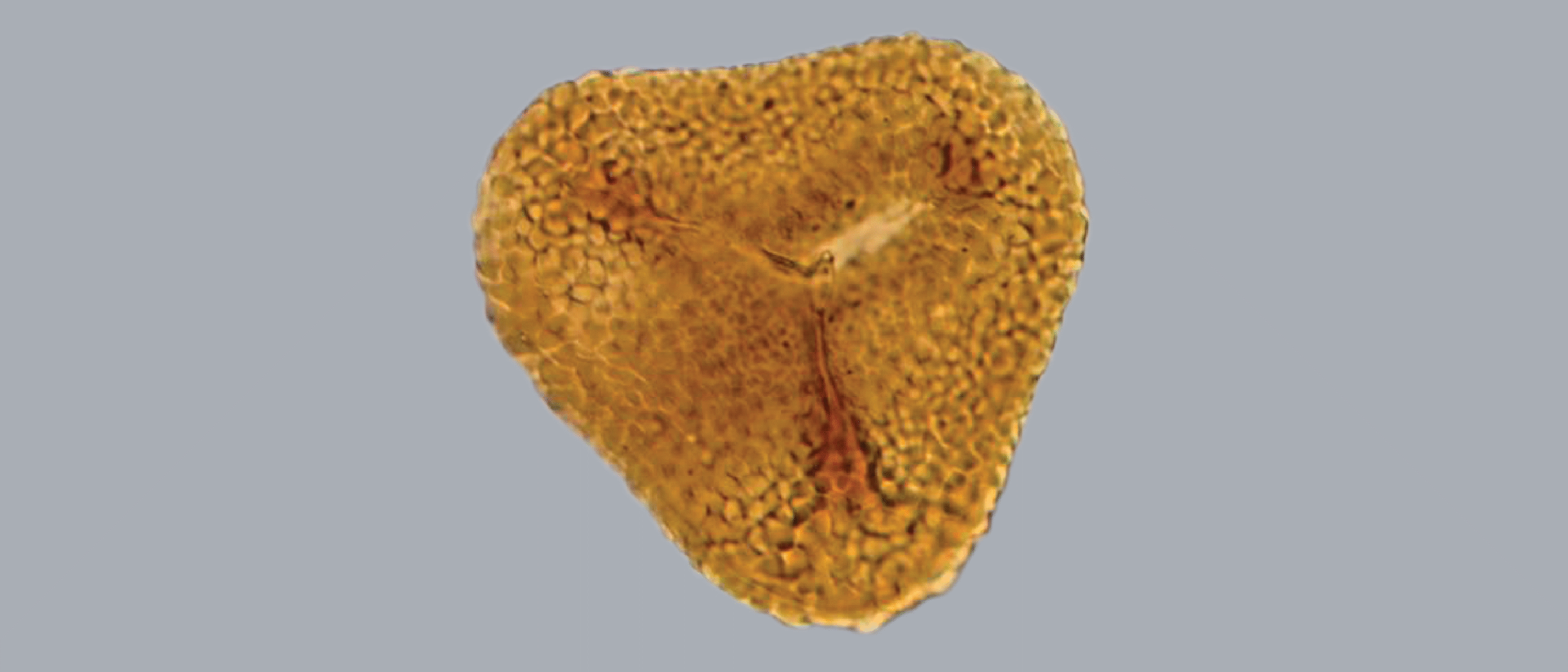
Palynologists are palaeontologists who specialise in microfossils composed of acid-resistant carbon-hydrogen-oxygen biopolymer. In other words, their fossils are nature’s microplastics. Resistant to almost anything diagenesis can throw at them, fossil microplankton, pollen and spores from the Earth’s deep past are sometimes more abundant and better preserved than their descendants deposited on the seabed today. These resilient fossils are therefore a major part of biostratigraphic analysis in petroleum exploration.
Biostratigraphy is a fundamental analytic tool for reducing economic risk by improving our understanding of palaeoenvironments, palaeogeography, and basin history.
At the upcoming Digex Conference – 28-30 May in Oslo – Robert Williams will present about the scanning project he is involved with at the NPD.
“Multi-gigapixel Scans of Fossil Microplankton, Pollen and Spores from Cores and Cuttings replace Microscopes with Desktops”
Like all fields of palaeontology, palynology is grounded on taxonomy – identifying tiny fossils to the species level based on differences in both gross and fine structure at sub-micron resolution. This science appeals to geologists who are skilled in recognising visual patterns and morphological minutiae among copious numbers of complex forms.
Palynologists enjoy painstaking, meticulous, detailed visual analysis through a microscope. They identify and count thousands of specimens of thousands of species recovered from thousands of well samples, and reduce this colossal complexity to three essentials: a sediment’s age of deposition, its environment of deposition, and the presence of eroded, re-deposited sediments.
Palynological analysis is slow. Optical design and imaging technology have greatly improved, but microscopy has remained essentially unchanged for four centuries. Microscopists in a biostratigraphy laboratory are limited to examining one slide at a time. Other users may have to wait for weeks or months for access to a set of released slides from one or more wells.
Because glass slides are thin and fragile, hundreds of NPD slides have been fractured or lost during shipment. Contrast this with seismic data or wireline logs which lend themselves well to simultaneous interpretation in different locations, simply because they are digital.
What an advantage it would be to have digital microscope slides in sub-micron resolution, instantly accessible by palynologists the world over!
Aerospace and pathology
Using an analogue charge-coupled device (CCD) sensor and video digitizer in the early 1990s, I experimented with imaging mosaics of analogue-to-digital photomicrographs for creating wide-field, high-resolution images of palynology slides. However, manual stitching proved too arduous to be effective as a routine method of documentation.
Image stitching was established methodology for pre-Apollo lunar reconnaissance. The Lunar Orbiter robotic missions of the mid 1960s used analogue photography to map the lunar surface. The robots chemically developed the film onboard the spacecraft. As if this wasn’t impressive enough, the Lunar Orbiters digitally scanned the developed negatives and transmitted them as binary data to Earth, where technicians stitched images together to make high-resolution mosaic prints. Amateur astronomers adopted similar techniques for astrophotography with homemade CCD cameras in the early 1990s, long before digital imaging was commonplace commercially or in palynology.
Unknown to me thirty years ago was that pathologists had already taken their first steps into the realm of digital whole slide imaging. Called telepathology, early trials of analogue electronic transmission of microscope slides were already improving diagnoses through interprofessional communication since the 1960s. In 1998, pathologists entered the digital realm with the advent of the first high-resolution whole slide scanners.
Replacing the words pathology with palynology, and tissue with palynomorphs, diagnosis with interpretation, every presentation was relevant for palynology’s digital future.
However, as late as 2011, whole slide scanners still did not attain sufficient speed and image resolution suitable for palynology. Perhaps this reflected the lesser optical resolution demands for pathology analysis, since pathology was the economic driver behind scanner technology.
After attending a slide scanner presentation in 2017, I and my palynology colleagues in Stavanger realised that whole slide scanners had finally achieved our required four pixel per micron resolution.
Fortunately, around the same time, the concept of digitalisation became prioritised in resource management at the NPD. Instead of simply making analogue data digital, digitalisation aimed to transform inefficient analogue processes into faster, automated digital processes.
With this goal in mind for palynology, the NPD sent me to The 14th European Congress on Digital Pathology, held in Helsinki in 2018. Every paper at this conference dealt with procedures, case histories and technology for the application of whole slide tissue scanning. Through machine learning and digital slide analysis among networks of medical specialists across national boundaries, the digital pathologists’ goal was to improve diagnosis of diseased tissue. Replacing the words pathology with palynology, and tissue with palynomorphs, diagnosis with interpretation, every presentation was relevant for palynology’s digital future.
This pathology conference made clear that we had to set in motion the digitisation of NPD’s 120 thousand palynology slides as soon as possible. In addition to NPD slides, there exists at least as many slides stored in companies’ and consultant laboratories’ slide archives. All must be digitised.
Avatara-p and Diskos
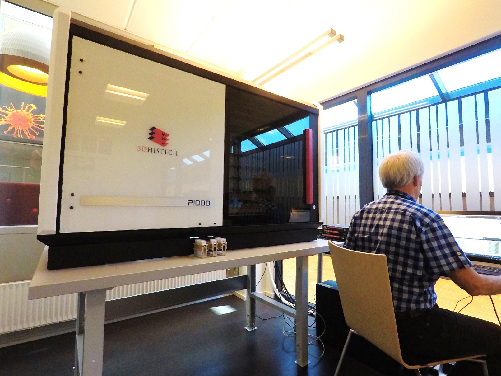
Within eight months after the Helsinki pathology conference, the NPD became the second institution in Europe to acquire the highest capacity, highest resolution whole slide scanner available, the 3DHistech P1000. The NPD’s scanner was the second P1000 in the production queue after 3DHistech delivered the first one to the General Hospital of Vienna, Austria in 2019.
The NPD’s digitalisation project Avatara-p – Advanced Augmented Analysis Robot for Palynology – had become a reality.
In spite of an eleven month scanner shutdown and maintenance delays during the Covid-19 pandemic, Avatara-p has now produced almost forty thousand digital palynology slides from approximately 300 exploration and development wells. Currently at 57 terabytes, the NPD is steadily expanding this dataset.
This entire dataset is now available on Diskos.
By interrogating these digital slides with machine learning applications trained and quality-controlled by palynology teams and laboratories, the re-analysis of microplankton, pollen and spores may reduce complexity even more.
Palynologists have increased our knowledge of great swaths of Norway’s deep history through fifty years of thorough analysis of uncounted numbers of palynology slides. Augmented analysis through machine-learning will potentially unearth even more unknown vistas in Norway’s deep history.